Nalvera.AI Cloud-AI platform for
medical imaging research

Upload a scan, select an AI task, and get results in your browser. No installation or programming required.

  • Easy-to-use AI models for CT and MRI
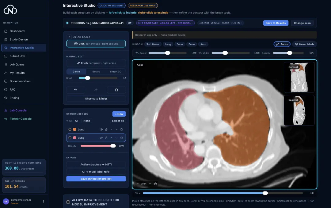
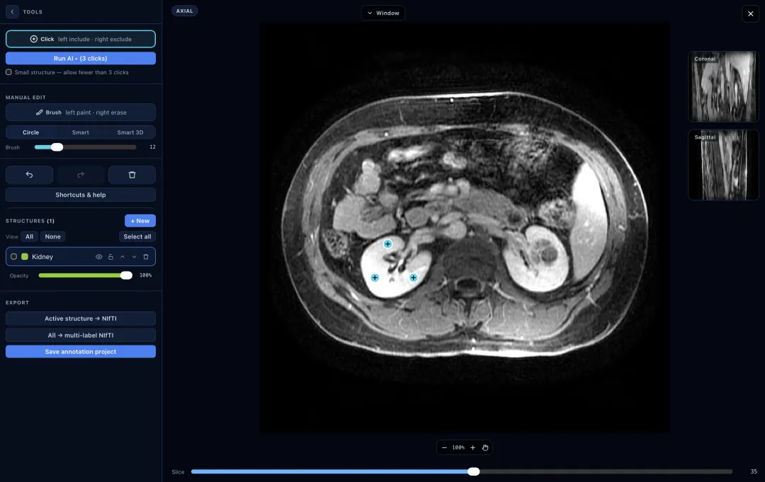
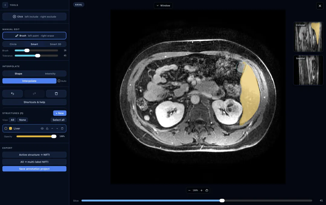
  • Click-to-segment Interactive AI Studio
  • Study Designs — follow patient measurements across scans
  • Export open medical-imaging formats
  • Your data stays yours in EU-hosted infrastructure
Run your first AI analysis
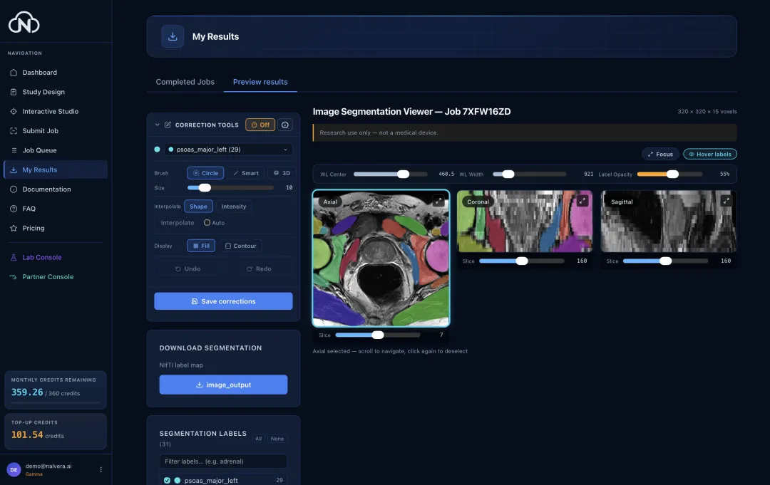
Product preview
Scroll
How it works

From scan to result in four steps

Upload your medical image

Drag and drop your scan. All common research imaging formats are supported.

Pick an AI model from our library

Choose from peer-reviewed and in-house models. You see the cost before you run.

Submit & monitor AI tasks

Your AI task runs in the cloud on our GPUs. Follow its progress live.

View, adapt, and download results

Review results in the browser, refine where needed, and download in standard formats.

Capabilities

Segment, generate, report — one workflow

Image segmentation

Label anatomy two ways

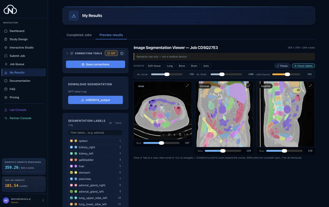
Guide the AI model click by click, or let it label every structure in one pass. Both run in the browser, where you can check and correct the result.

Nalvera iSeg · in-house TotalSegmentator MOOSE MuscleMap CT / MR
A brain MR with the cerebellum outlined in orange by the click-guided AI.
Output · brain MR · cerebellum
Image generation

Create synthetic scans from scratch

Create a CT or brain MR scan of a patient who does not exist. Choose the body region and the image size, then download it like any other AI task. Useful for enlarging a small dataset, demoing software, or sharing an example without real patient data.

MAISI-v2 No input scan needed
A synthetic thorax CT slice, generated by AI and shown at lung window settings.
Output · thorax 3D CT
A synthetic T2-weighted brain MR slice, generated by AI.
Output · brain T2 MR
Image captioning

From scan to written report

Merlin reads a whole abdominal CT and writes a structured report, one section per organ system. It can also rank the most likely findings, or estimate five-year risk for six chronic conditions.

Merlin · vision-language AI 13 organ systems Reports · findings · risk
AI task Merlin · Radiology report
The input to the captioning example: an axial CT slice through the mid-abdomen showing both kidneys, the aorta, the vertebral body and gas-filled bowel loops, at soft-tissue window settings. Input · abdominal 3D CT
Merlin output
[kidneys] The kidneys enhance homogeneously. No focal renal lesions. No hydronephrosis or hydroureter involving either collecting system. [bowel] There is no evidence of bowel obstruction. [circulatory] Atherosclerosis without aneurysm. [musculoskeletal] No suspicious osseous lesions identified. [peritoneum] No free fluid. Abdominal wall: normal. [lower thorax] No significant pulmonary, pleural, or mediastinal abnormality is seen in the visualized portions of the thorax. [liver] Normal in size. There are multiple subcentimeter hypodensities throughout both lobes of the liver, which likely represent cysts or hamartomas but are too small to characterize on this examination. No intrahepatic or extra hepatic ductal dilatation. [gallbladder] Surgically absent. [pelvic] Normal. Prostate and seminal vesicles: the prostate is mildly enlarged measuring 5 cm in transverse dimension (3/401). [lymph nodes] No enlarged retroperitoneal, mesenteric, pelvic or inguinal lymph nodes. [spleen] Normal. [pancreas] Normal. [adrenal glands] Normal.

Run your first scan today

Free to start, right in your browser. No installation, no card required.